Jeff Lichtman
Series 3 Episode 3
The Society of Cells: Brainbow Mapping with Jeff Lichtman
Series 3 Episode 3
Available on all major podcast platforms: click on the icons below or on 'play' to listen now.
In this episode of What's Your Map?, neuroscientist and Harvard Professor Jeff Lichtman walks Jerry through his astonishing work mapping the human brain. In 2024, with the help of Google Research, Jeff’s team developed the most detailed map of a brain sample ever created, producing 1,400 TB of data from a sample the size of a pinhead.
Jeff also talks about how he and Harvard biologist Dr Joshua Sanes pioneered the Brainbow process, a breakthrough that allows scientists to easily identify individual neurons in the brain by colour.
He explains how human experiences allow us to build neural maps, or wiring diagrams, onto our brains. These maps encode our memories and learned experiences, so that we can act without thinking, like riding a bicycle. However, they do not always work as they should.
Zoom in on this extraordinary image as Jerry and Jeff discuss the map:
Map of Large and Small Neurons © Google Research & Lichtman Lab / Harvard University
This extraordinary image may look like an artwork or a faraway galaxy - but it is in fact inner space: a tiny fragment of a human brain!
The sample used for this Brainbow map was removed from the hippocampus of a brain during neurosurgery, in an attempt to correct epilepsy. The hippocampus is responsible for organising and storing memories, and for spatial navigation.
This ‘chunk’, as Jeff describes, was just one cubic millimetre - but still big enough to contain 57,000 cells, and 150 million synapses! It was sliced incredibly thinly and photographed using a multibeam scanning electron microscope. Aided by Google Research, the images were then aligned and compiled to create a vast wiring diagram of nerve cells, their connections, and functions.
This illustration shows a signal working its way down an axon to the cell body and dendrites of the next cell. By illustrator: Christy Krames, MA, CMI, for US National Institutes of Health, National Institute on Aging - Public Domain, via Wikimedia Commons.
The human brain is composed of approximately 86 billion nerve cells, or neurons.
Each neuron is made up of three general components; a cell body or ‘soma’ that contains the nucleus which controls the cell's activities. Out of this cell body emerges an axon, a tendril-like extension which acts as a vessel to send information to other cells by electrical impulse.
At the ends of the axon are branches known as synapses, where the axon releases chemicals onto the dendrites of other neurons.
Dendrites are where neurons receive synaptic connections from the axons of other cells, and each cell has one axon that makes a connection with another.
This creates a continuous network of connections, meaning that information can flow like a news broadcast through these cells. Jeff has likened the axon as how one talks to and transmits information, and the dendrites as ears or eyes and how one receives information.
Public Domain Photograph by Simi Iluyomade on Unsplash
When uncoloured, Jeff explains that a map of the brain can look similar to that of an abstract expressionist painting by the likes of Jackson Pollock - where there appears to be no rhyme or reason.
The viewer's understanding is transformed as soon as colour is introduced!
The image has been created using the pioneering Brainbow technique, where scientists colour or stain neurons using fluorescent proteins. This allows each neuron to be labelled individually, highlighting pathways of connectivity, tracing synapses, and distinguishing their size. You can find out more about this ground-breaking method here.
In this technicolour snapshot of a mammalian brain, the cells are colour-coded according to size - the smallest in blue to the largest cells in red. The resulting image shows how the brain cells are organised into 6 cortical layers, separated by type and density:
Cropped image of large and small neurons to highlight cortical layering © Google Research & Lichtman Lab / Harvard University
The small blue/green cells make up the molecular layer. This is the most superficial, containing only a few neurons. It also features glial cells, a type of cell that helps to support neurons.
The orange/yellow cells are the external granular and extra pyramidal layers. They are often combined and contain small to midsize pyramidal cells and local circuit neurons.
Pyramidal cells, named so because of their shape, are vital for processing senses like sight, touch, and sound, and for motor control (planning and executing movements). They also play a role in complex processes like learning and decision-making.
A second blue layer makes up the internal granular layer, which receives input from the brain's sensory relay station, the thalamus. The thalamus in turn gets information from other parts of the brain.
The next, internal pyramidal layer, contains large red cells and is a major output of data.
Finally, the multiform layer transmits information to the thalamus and other cortical areas. It contains a diverse population of neurons.
Not shown here in this image is the white matter, where axon cables send their information to other parts of the brain.
The Brainbow method was developed in 2007 by Professors of Molecular & Cellular Biology Jeff W. Lichtman and Joshua R. Sanes, Jean Livet, and their team at the Department of Neurobiology at Harvard University.
They collaboratively published a paper on their work in NATURE magazine: "Transgenic strategies for combinatorial expression of fluorescent proteins in the nervous system". You can find this paper here.
The mouse brain and the human brain have many similarities, including that a mouse has 75 thousand neurons, and a human has 86 thousand. Therefore, mice were the first organisms that this neuroimaging technique was successfully tried on.
Here are three Brainbow maps of mouse neurons by Lichtman and Sanes (2008):
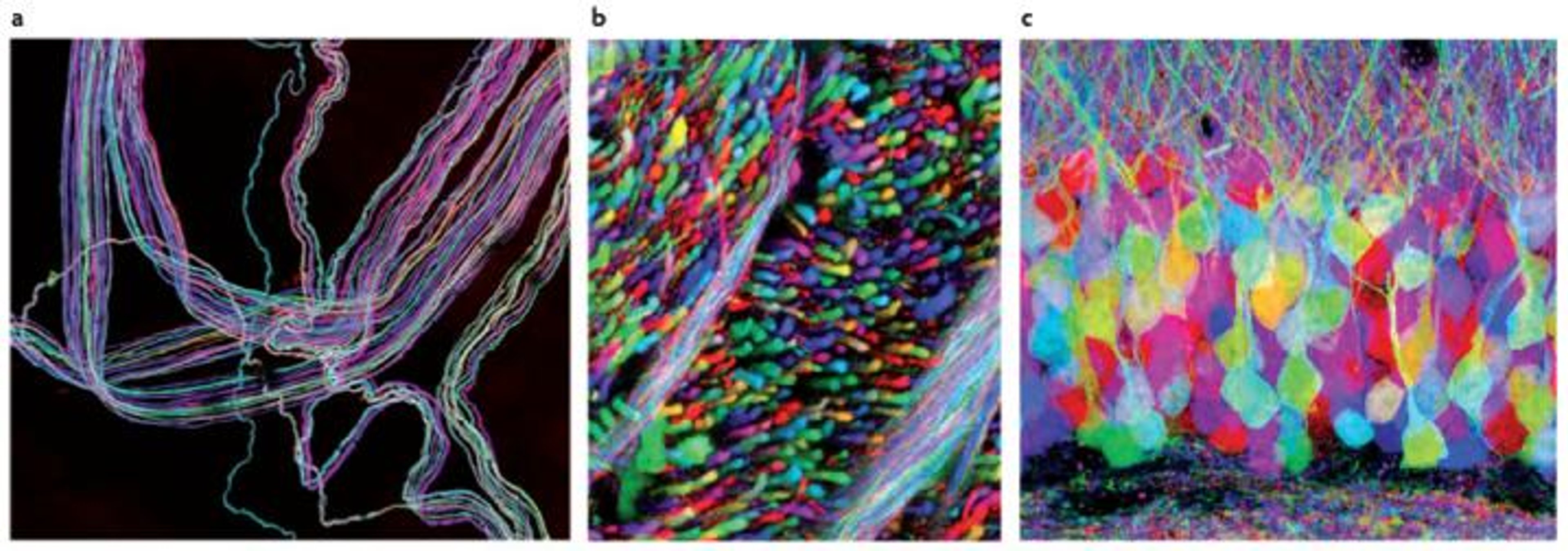
Fig. a: A motor nerve supplying an ear muscle, Fig. b. An axon tract in the brain stem, Fig. c. The hippocampal dentate gyrus [a part of the brain's hippocampal formation, playing a critical role in memory formation and spatial navigation. Credit: National Library of Medicine. CC BY 3.0, via Wikimedia Commons.
Another mind-blowing image from the Lichtman Lab: A single neuron with its dendrites, showing the thousands of axons connecting to it:
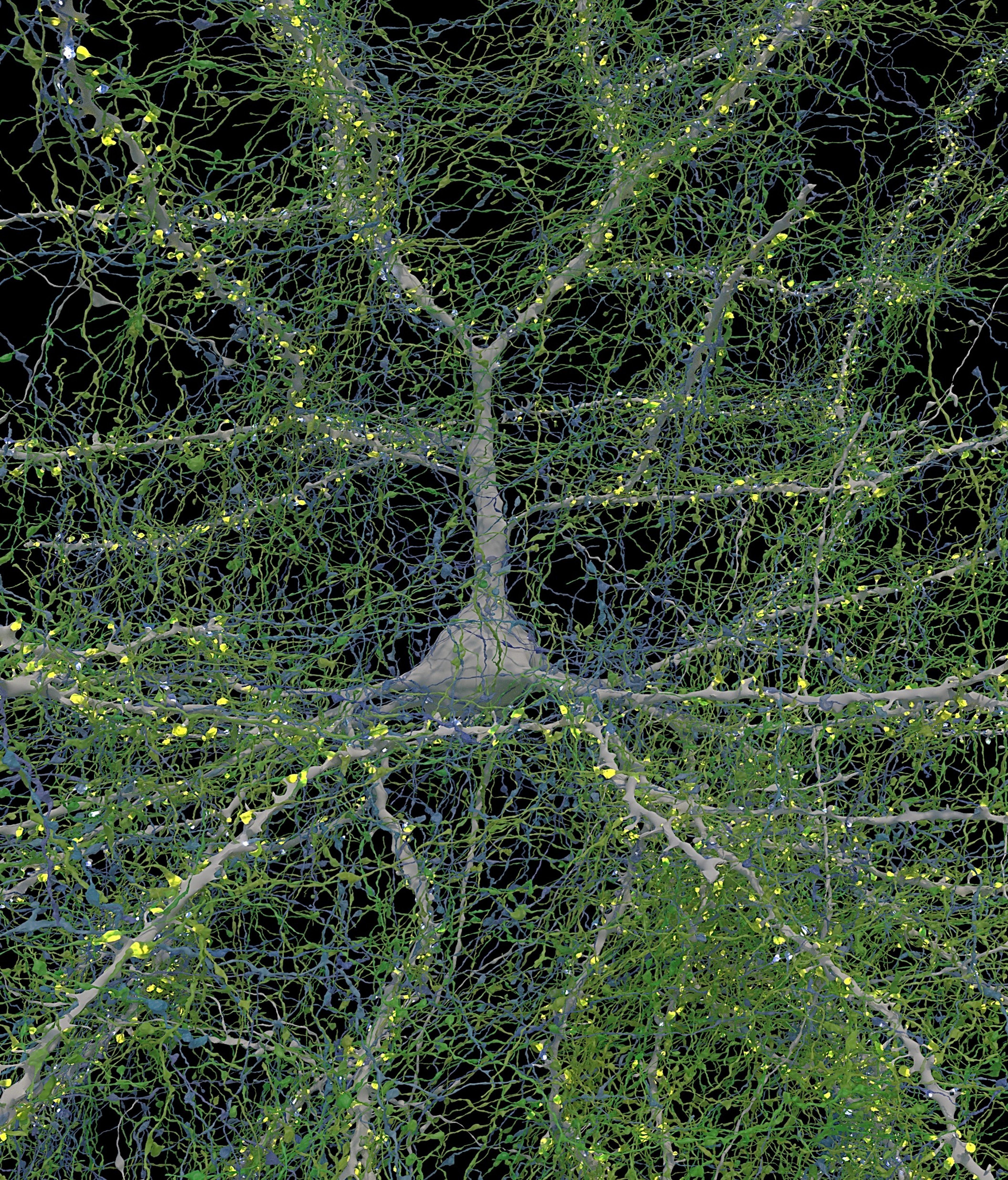
Image of a single neuron © Google Research & Lichtman Lab/Harvard University
In 2024 during a collaboration with Google Research, the Lichtman lab produced the most detailed map of a human brain sample ever made. You can read more about this here on the Harvard University Gazette.
Being able to map the brain is no mean feat. It is the result of generations of scientists working in collaboration, building on research and investigation, and in fact the trailblazing Brainbow technique finds its foundations in the 19th century...
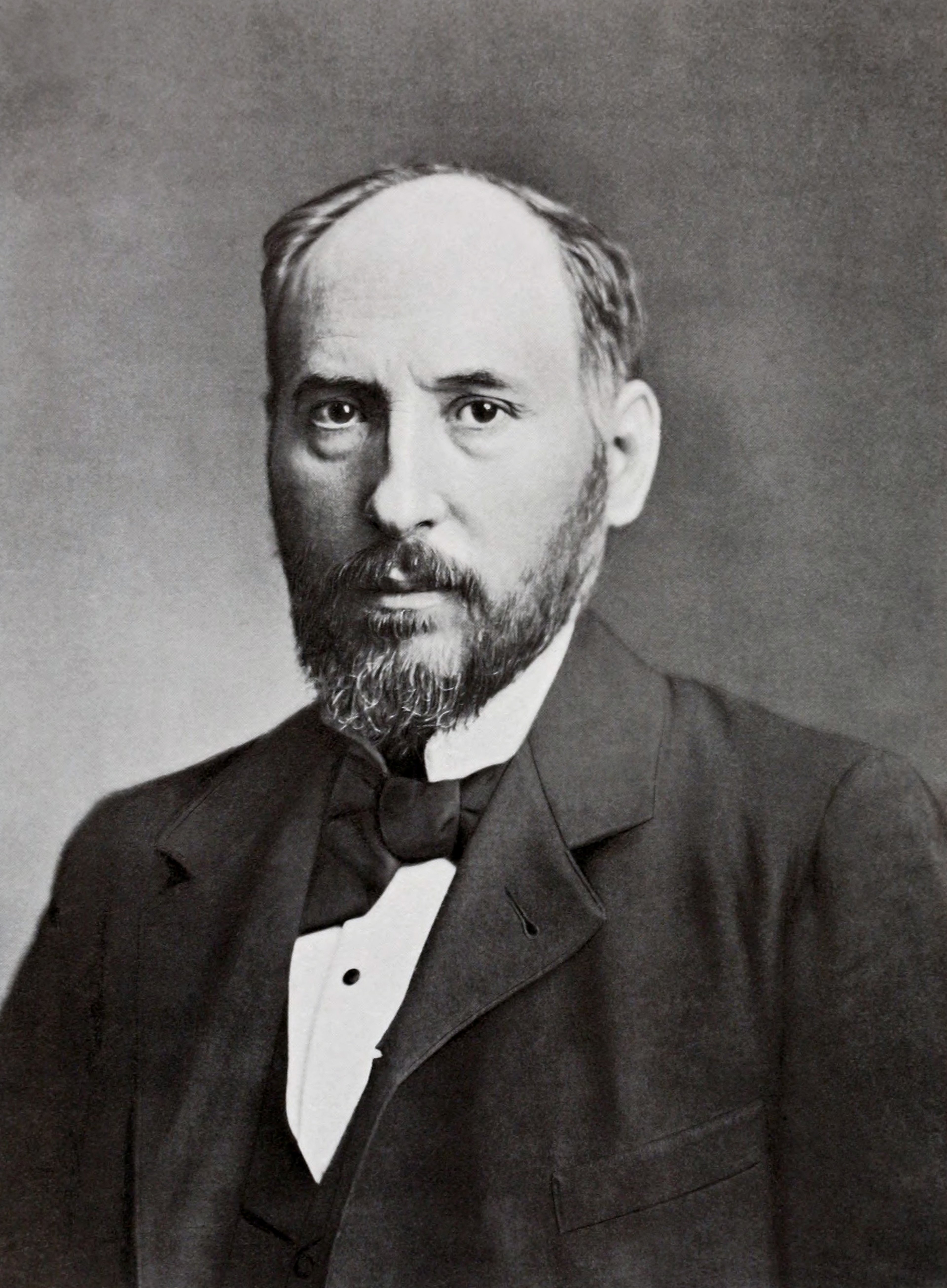
Santiago Ramón y Cajal. Published by Clark University in 1899. Public Domain, via Wikimedia Commons.
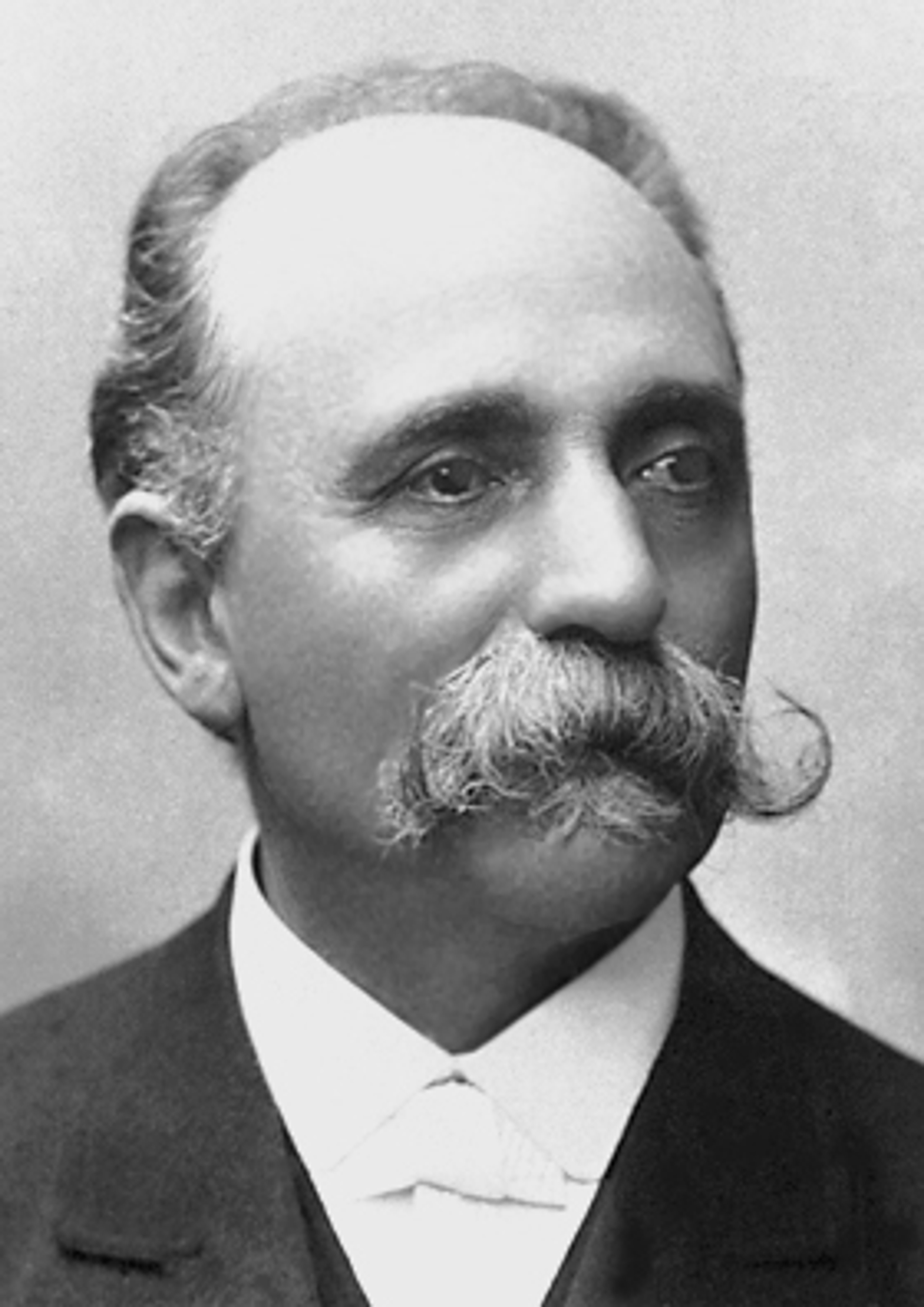
Camilo Golgi, Unknown author. Public Domain, via Wikimedia Commons
Santiago Ramón y Cajal (1852-1934) was a Spanish scientist. In 1887 while working as a Professor in Barcelona, he learnt of a method by Italian pathologist, Camilo Golgi (1843-1926), which involved staining cells with a silver chromate solution - now known as the Golgi Stain.
This ‘black reaction’ as it was known, was a revolutionary technique that selectively stained a small amount of nerve cells, and when examined under a microscope, revealed their intricate structures.
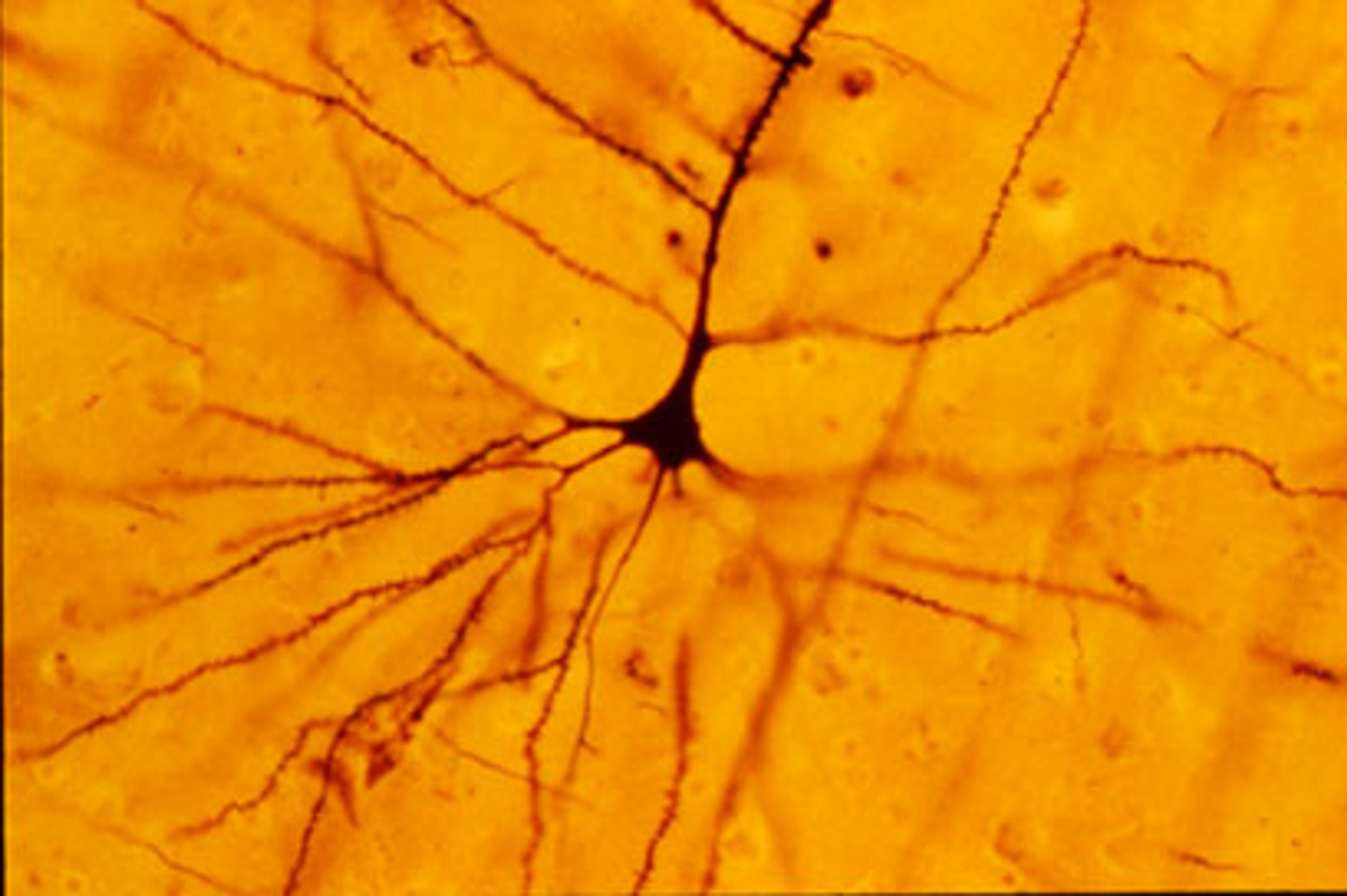
Credit: Bob Jacobs, Colorado College. By Cahass. CC BY-SA 3.0.
This image shows a human pyramidal neuron from the neocortical layer of the brain (an outer layer connected with higher thinking and intelligence) stained using the Golgi Technique.
Ramón y Cajal employed Golgi's staining method to examine the nervous system in incredible detail, paying particular attention to the brain and spinal cord. His keen observations were paired with his skill as a draughtsman: Cajal created incredible sketches of neurons and cell structures.
Similar to the imagery produced by the Lichtman Lab, these drawings of cortical layering by Cajal show a vertical cross-section, with the surface of the cortex at the top (some using Golgi staining method and other using a staining method by German neuroscientist Franz Nissl):
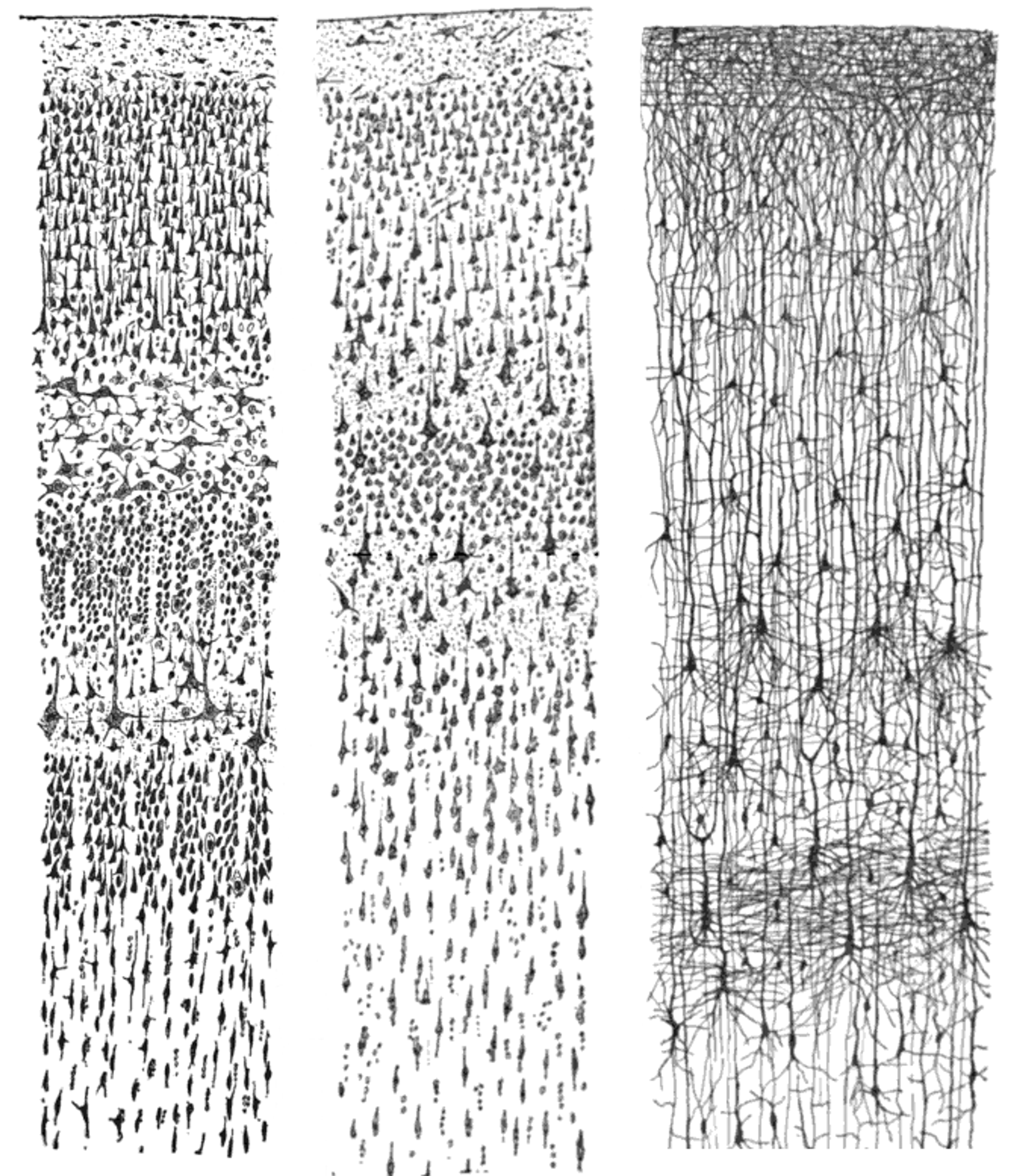
Credit: Looie496. Public Domain, via Wikimedia Commons.
From left: Nissl-stained visual cortex of a human adult. Middle: Nissl-stained motor cortex of a human adult. Right: Golgi-stained cortex of a 1+1⁄2 month-old infant. The Nissl stain shows the cell bodies of neurons; the Golgi stain shows the dendrites and axons of a random subset of neurons.
Originating from "Comparative study of the sensory areas of the human cortex" by Santiago Ramón y Cajal, published in 1899.
His work led him to make fundamental discoveries that the nervous system is made up of individual neurons, rather than a continuous network!
In 1906, Ramón y Cajal and Golgi were jointly awarded a Nobel Prize in Physiology or Medicine for their work, which formed the foundation for our understanding of the nervous system. Cajal is now known as the ‘Father of Neuroscience’.
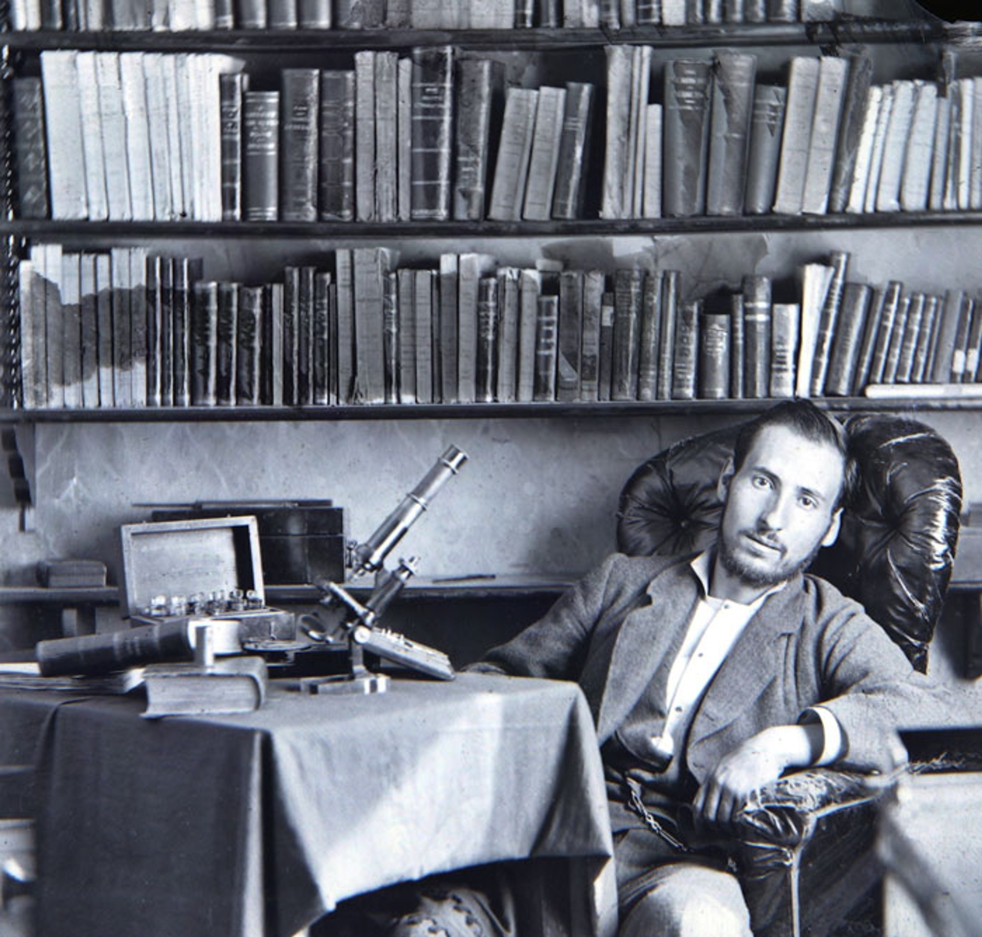
Self-portrait as a student, c.1870. By Santiago Ramón y Cajal. Public Domain.
During the podcast, Jeff introduces us to the world of Connectomics, a field of neuroscience focused on mapping the connections within a nervous system. These maps, or connectomes, illustrate the structure and organisation of the brain, and facilitate the study of the connections and behaviours of the individual neurons.
Connectomes also enable the study of the structure and changes in the brain throughout the course of a person's life. They are particularly useful in researching brain disorders such as Alzheimer's or epilepsy. Often the physical structure indicates the medical issue, and scientists have to work backwards to identify the cause. Connectomics can help to provide valuable insights.
And, when it comes to learning in the field of neuroscience, Jeff acknowledges how data is now much more easily and quickly produced and processed due to technology and machine learning, which has transformed the field. Data sets are generated and processed so fast that he and his colleagues struggle to keep up!
Jeff has spent his career studying how experiences are encoded into the brain. As he explains, animals are born with experiences and reflexes, while humans know much less when they enter the world, and must learn everything in order to survive. Since children have much more elastic brains than adults, they can record more information and pick up skills and reflexes faster than an older or aging brain.
And, as Jerry knows all too well, learning how to ride a bike as an adult is much harder than it would be for a child. It is also something that can only be learned through the physical experience of trial and error. This eventually becomes ‘procedural memory’, almost a subconscious reflex, by creating a neural pathway that allows the brain to send complex information for the body to complete a task. This is integrated with sensory information, such as learning to balance.

Credit: Alextredz - Own work, CC BY-SA 4.0
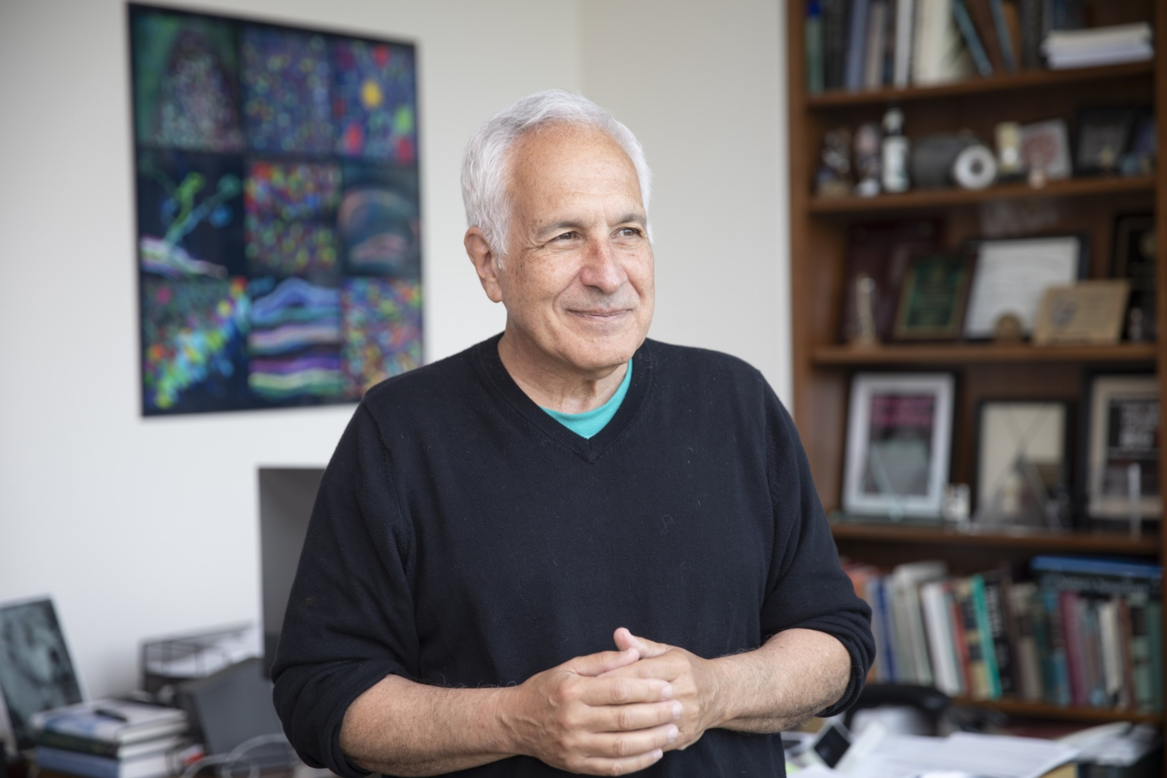
About Jeff Lichtman

Jeff Lichtman is Jeremy R. Knowles Professor of Molecular and Cellular Biology at Harvard University.
He received an AB from Bowdoin (1973), and an M.D. and Ph.D. from Washington University (1980) where he worked for 30 years before moving to Cambridge, MA, in 2004. He is a member of the Center for Brain Science.
Lichtman’s research interest revolves around the question of how mammalian brain circuits are physically altered by experiences, especially in early life. He has focused on the dramatic re-wiring of neural connections that takes place in early postnatal development when animals are doing most of their learning.
This work has required development of techniques such as Brainbow mapping of mice to visualize neural connections and monitor how they are altered over time.
Recently, his efforts have focused on developing new electron microscopy methods to map the entire wiring diagram of the developing and adult brain. This connectomics-based approach aims at uncovering the ways information is stored in neural networks.
You can catch up on the lectures delivered by Jeff here.
A new episode of WHAT'S YOUR MAP? will be available every two weeks. Why not continue your exploration of the wonderful world of maps by subscribing? That way you'll never miss an episode.
Feel free to let us know - What's YOUR Map?!

